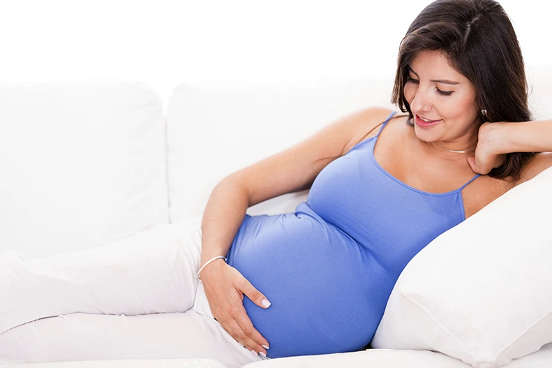
New York [US]: A team of anthropologists observed that replica completely changes the bones of females in approaches that had been previously unknown. Its revelation, based totally on primatological research, offers clean light on how giving birth may permanently regulate the frame.
The findings of the observation had been posted in the magazine PLOS ONE. “Our findings offer additional proof of the profound effect that reproduction has on the lady organism, similarly demonstrating that the skeleton isn’t a static organ, however a dynamic one which modifications with life events,” explains Paola Cerrito, who led the studies as a doctoral scholar in NYU’s Department of Anthropology and College of Dentistry.
Specifically, the researchers discovered that calcium, magnesium, and phosphorus concentrations are decreased in girls with skilled reproduction. These changes are related to giving delivery itself and to lactation.
However, they caution that while other clinical research show calcium and phosphorus are essential for foremost bone energy, the new findings do not address normal health implications for either primates or humans. Rather, they are saying, the work illuminates the dynamic nature of our bones.
“A bone isn’t always a static and dead portion of the skeleton,” notes NYU anthropologist Shara Bailey, one of the examine’s authors. “It continuously adjusts and responds to physiological methods.”
The different authors of the take a look at, which appears within the magazine PLOS ONE, are Timothy Bromage, a professor in NYU College of Dentistry, Bin Hu, an adjunct professor at NYU College of Dentistry, Justin Goldstein, a doctoral candidate at Texas State University, and Rachel Kalisher, a doctoral candidate at Brown University.
It’s long been installed that menopause could impact females’ bones. Less clear is how preceding lifestyles-cycle events, which include duplicate, can impact skeletal composition. To cope with this, the researchers studied the primary lamellar bone–the main form of bone in a mature skeleton. This element of the skeleton is an excellent part of the frame to examine because it changes over time and leaves biological markers of those adjustments, permitting scientists to screen alterations throughout the existence span.
In the researchers examined the growth rate of lamellar bone within the femora, or thigh bones, of each girl and male primates who had lived at the Sabana Seca Field Station in Puerto Rico and died of natural causes.
Veterinarians at the field station monitored and recorded records on those primates’ fitness and reproductive records, allowing the researchers to in shape bone-composition changes to existing events with amazing precision.
Cerrito and her colleagues used electron microscopy and strength-dispersive X-ray evaluation–commonly deployed methods to gauge the chemical composition of tissue samples–to calculate changes in calcium, phosphorus, oxygen, magnesium, and sodium concentrations in the primates’ bones.
Their effects confirmed distinct concentrations of some of these factors in females who gave start compared men in addition to women who did now not deliver birth. Specifically, in women who gave beginning, calcium and phosphorus were lower in bone fashioned at some stage in reproductive occasions. Moreover, there was a extensive decline in magnesium concentration throughout these primates’ breastfeeding of toddlers.
“Our studies suggest that even before the cessation of fertility the skeleton responds dynamically to changes in reproductive reputation,” says Cerrito, now a research fellow at ETH Zurich. “Moreover, those findings reaffirm the great impact giving start has on a girl organism–quite genuinely, evidence of reproduction is ‘written within the bones’ for life.”